
Mitochondrial Dysfunction: Symptoms, Causes, Testing & Cellular Energy
Mitochondria produce most of the ATP that powers our cells, making healthy mitochondrial function especially important in high-energy tissues such as the brain, muscles, heart, kidneys and retina. When mitochondrial energy production becomes impaired, possible symptoms can include fatigue, exercise intolerance, muscle weakness, poor recovery, brain fog, cognitive changes and visual or neurological problems.
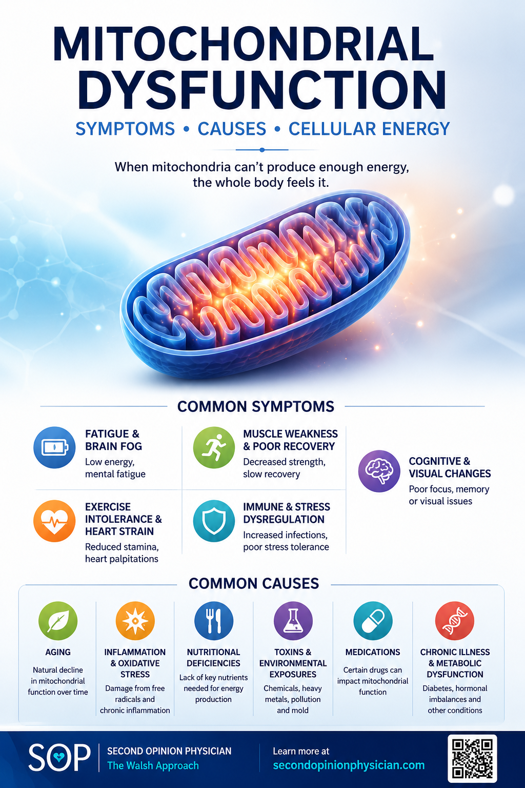
Mitochondrial dysfunction does not necessarily mean a rare inherited mitochondrial disease. It may also develop or worsen with aging, oxidative stress, chronic inflammation, metabolic dysfunction, nutritional deficiencies, medications, toxins and chronic illness. Because there is no single test that identifies every mitochondrial problem, evaluation often looks at a pattern of symptoms together with markers such as GDF-15, lactate, pyruvate, creatine kinase, carnitine and acylcarnitines to better understand cellular energy metabolism and determine what may be driving the dysfunction.
Mitochondria produce most of the usable cellular energy required by the brain, muscles, heart, retina and other high-demand tissues. When mitochondrial function becomes impaired, the effects can extend well beyond fatigue—affecting exercise tolerance, cognition, muscle function, recovery and metabolic resilience.
But mitochondrial dysfunction is not one disease or one biochemical problem. It may result from an inherited mitochondrial disorder or develop secondarily from nutrient deficiencies, oxidative stress, inflammation, metabolic dysfunction, medications, toxic exposures or other physiological stressors.
For some patients, mitochondrial stress also overlaps with methylation and epigenetic regulation. This is particularly relevant to our expanded approach to undermethylation, where mitochondrial distress may represent one of several biochemical drivers contributing to impaired methylation rather than an isolated diagnosis.
Key clinical concept: There is no single test for mitochondrial dysfunction. The goal is to identify the clinical pattern, examine appropriate markers of cellular-energy metabolism, and then determine what may be driving the dysfunction.
In This Guide
What Are Mitochondria and Why Are They Important?
Mitochondria are specialized structures within most human cells. Their best-known role is converting energy from carbohydrates, fats and proteins into adenosine triphosphate (ATP).
ATP is the immediately usable energy required for muscle contraction, nerve signaling, membrane transport, cellular repair, protein synthesis and thousands of other biological processes.
Inside the inner mitochondrial membrane is the electron transport chain. Electrons derived from nutrients move through a series of protein complexes, creating the electrochemical gradient used to manufacture ATP.
Mitochondria convert energy from food into ATP that cells can immediately use.
Mitochondria participate in carbohydrate, fatty-acid and amino-acid metabolism and influence cellular redox balance.
Mitochondria influence oxidative signaling, calcium handling, stress responses and programmed cell death.
What Is Mitochondrial Dysfunction?
Mitochondrial dysfunction occurs when mitochondria cannot perform their normal cellular functions efficiently. The problem may involve ATP production, electron transport, mitochondrial membranes, excessive oxidative stress, mitochondrial turnover or several processes simultaneously.
This does not necessarily mean complete mitochondrial failure. A person may instead have a reduced ability to increase cellular energy production when demand rises.
The body may produce adequate energy at rest but have difficulty meeting increased energy demands during exercise, illness, metabolic stress or prolonged cognitive activity.
What Are the Symptoms of Mitochondrial Dysfunction?
There is no single symptom that identifies mitochondrial dysfunction. Symptoms depend upon the cause, severity and which tissues are affected.
Fatigue, reduced endurance, exercise intolerance and unusually prolonged recovery after activity.
Muscle weakness, aching, early fatigue or difficulty sustaining activity.
Brain fog, cognitive difficulty and neurological symptoms in some mitochondrial disorders.
Retinal, optic or hearing abnormalities occur in several inherited mitochondrial conditions.
Some mitochondrial diseases affect cardiac muscle or electrical conduction because of the heart's enormous ATP requirement.
Combined muscle, neurological, metabolic, cardiac, visual or hearing findings can increase suspicion of mitochondrial disease.
Why Are the Brain, Muscle, Heart and Retina So Vulnerable?
Tissues that require large and continuous amounts of ATP are especially dependent upon mitochondrial performance.
Neurons continuously use ATP to maintain membrane potentials, transmit signals, recycle neurotransmitters and preserve cellular structures.
Energy demand rises dramatically during activity, which is why mitochondrial disorders frequently produce poor endurance and exercise intolerance.
The heart contracts continuously and depends heavily upon oxidative metabolism and mitochondrial ATP.
Photoreceptors and retinal pigment epithelial cells have extraordinary metabolic demands, making mitochondrial health particularly relevant to retinal function.
Renal tubular cells use large amounts of ATP for continuous membrane transport and therefore contain abundant mitochondria.
Long neurons require substantial energy for axonal transport and maintenance, making neurological symptoms possible in mitochondrial disorders.

Primary Mitochondrial Disease vs. Acquired Mitochondrial Dysfunction
This distinction is essential because not every patient with evidence of mitochondrial stress has an inherited mitochondrial disorder.
Primary Mitochondrial Disease
Primary mitochondrial diseases result from pathogenic genetic variants affecting mitochondrial structure or function.
The abnormality may involve mitochondrial DNA or nuclear DNA containing genes required for normal mitochondrial function.
When primary mitochondrial disease is suspected, biochemical testing may provide clues, but genetic evaluation is often required.
Secondary / Acquired Dysfunction
Mitochondrial performance can also become impaired as a consequence of another physiological problem.
Potential contributors include metabolic dysfunction, nutrient deficiencies, inflammation, oxidative stress, medications, toxins, chronic illness and aging.
Here mitochondrial dysfunction may be part of the disease process rather than the original cause.
What Can Cause Acquired Mitochondrial Dysfunction?
Acquired mitochondrial dysfunction can develop through several different pathways. Identifying the dominant contributor may be more useful than simply adding another mitochondrial supplement.
Mitochondrial enzymes require vitamins, minerals and metabolic cofactors to function normally.
Excess reactive oxygen species can damage mitochondrial membranes, proteins and DNA.
Chronic inflammatory signaling can alter energy metabolism and increase cellular oxidative stress.
Insulin resistance and abnormal substrate metabolism can affect mitochondrial energy utilization.
Certain medications and environmental or metabolic exposures can impair mitochondrial pathways.
Mitochondrial quality control, membrane integrity and energy production may become less efficient with age or prolonged physiological stress.
Mitochondrial Dysfunction, Undermethylation & Epigenetics
Mitochondrial function and methylation should not be viewed as completely separate biochemical systems. Cellular energy, oxidative stress, antioxidant defense, amino-acid metabolism and methylation continually interact.
This becomes particularly relevant in patients with an undermethylation pattern. The traditional Walsh Approach uses characteristic symptoms and biochemical markers to help identify undermethylation and other biotypes. Our expanded approach asks an additional question:
Rather than assuming that every undermethylated patient has the same biochemical problem, the Five Epigenetic Biotypes of Undermethylation examine several patterns that may contribute to impaired methylation and epigenetic regulation.
Different Biochemical Roads Can Lead to Impaired Methylation
Mitochondrial distress is one possible contributor. Other patterns may involve excessive methyl demand from creatine synthesis, oxidative or inflammatory stress, toxic/metabolic burden with impaired methylation flow, or other abnormalities affecting methyl-group availability and utilization.
Impaired cellular-energy production and altered redox biology may contribute to a biochemical environment that affects methylation and cellular regulation.
Endogenous creatine synthesis consumes SAM-derived methyl groups, creating a direct connection between cellular-energy biology and methylation demand.
Oxidative and inflammatory stress can affect mitochondrial function while also changing the biochemical environment surrounding methylation and gene regulation.
Accumulation of SAH can inhibit methyltransferase activity even when methyl-group supply appears adequate, producing functional methylation impairment.
How Should Mitochondrial Dysfunction Be Tested?
There is no single routine laboratory test that definitively diagnoses mitochondrial dysfunction. A practical evaluation uses several markers that examine different components of cellular-energy metabolism.
A cellular stress-response marker that can be elevated in primary mitochondrial disease and other conditions.
Provides information about the balance between glycolysis and mitochondrial oxidative metabolism.
Helps interpret lactate and provides additional information about glucose and cellular redox metabolism.
Helps identify skeletal-muscle injury or involvement but is not itself a direct mitochondrial function test.
Evaluates carnitine availability required for transport of long-chain fatty acids into mitochondria.
Examines fatty-acid oxidation and metabolic intermediates associated with several inherited energy disorders.
What Is GDF-15?
Growth Differentiation Factor 15 (GDF-15) is a stress-response protein that has emerged as an important biomarker in the evaluation of mitochondrial disease.
Mitochondrial dysfunction can activate cellular stress-response pathways that increase circulating GDF-15. It can be particularly useful when considered alongside the clinical phenotype and other metabolic testing.
Why Test Lactate and Pyruvate?
Glucose metabolism produces pyruvate. Under aerobic conditions, pyruvate can enter mitochondrial pathways that ultimately support ATP production.
When oxidative metabolism cannot adequately accommodate metabolic demand, more pyruvate may be converted to lactate.
↓
PYRUVATE
↙ ↘
LACTATE MITOCHONDRIA → ATP
Normal lactate does not exclude mitochondrial dysfunction. Lactate may be intermittently abnormal, and some mitochondrial disorders primarily affect specific tissues without substantially increasing circulating lactate.
The lactate-to-pyruvate relationship can sometimes provide additional information, particularly when lactate itself is elevated.
Why Test Carnitine and Acylcarnitines?
Long-chain fatty acids require the carnitine shuttle to enter mitochondria for beta-oxidation.
Helps determine whether adequate carnitine is available for fatty-acid transport and metabolism.
Looks for patterns of fatty-acid intermediates that can identify abnormalities in fatty-acid oxidation and certain organic-acid disorders.
What Can Urine Organic Acids Add?
Urine organic-acid testing measures metabolic intermediates generated through carbohydrate, amino-acid and fatty-acid metabolism.
Characteristic patterns can provide clues to organic acidemias, fatty-acid oxidation disorders and some abnormalities associated with mitochondrial respiratory-chain function.
Can Routine Blood Tests Be Normal?
Yes. A normal CBC, comprehensive metabolic panel, CK and even normal lactate do not exclude mitochondrial disease.
Some abnormalities emerge primarily during physiological stress, while others are concentrated within specific tissues.
This is why the clinical phenotype remains important. Striking exercise intolerance, muscle symptoms, neurological findings, retinal abnormalities or multisystem disease may justify further investigation despite relatively normal routine laboratory results.
Why Is Cardiolipin Important?
Cardiolipin is a specialized phospholipid concentrated in the inner mitochondrial membrane. It helps maintain membrane architecture and supports proteins involved in oxidative phosphorylation.
Damage or abnormal remodeling of cardiolipin can therefore interfere with mitochondrial energy production.
TAZ gene mutations disrupt normal cardiolipin remodeling, producing a primary mitochondrial disorder that commonly affects skeletal and cardiac muscle.
SS-31 is a mitochondria-targeted peptide that interacts with cardiolipin and the inner mitochondrial membrane rather than simply supplying an energy substrate.
Creatine, ATP and the Mitochondrial Energy Buffer
Mitochondria produce ATP, but cells also need a way to rapidly deliver and regenerate ATP when energy demand changes.
The creatine-phosphocreatine system provides this energy buffer. Mitochondrial ATP can transfer a phosphate group to creatine, forming phosphocreatine. Phosphocreatine can then rapidly regenerate ATP where it is needed.
This is particularly important in high-energy tissues such as skeletal muscle and brain.
Creatine & Mitochondrial EnergyHow Are Mitochondrial Function, Creatine and Methylation Connected?
One of the clearest biochemical connections between cellular-energy metabolism and methylation involves creatine synthesis.
Endogenous creatine production consumes methyl groups from S-adenosylmethionine (SAM) and produces S-adenosylhomocysteine (SAH). This means creatine synthesis contributes directly to methyl-group demand.
Mitochondrial energy production supports cellular metabolism, repair and biochemical reactions.
The phosphocreatine system buffers ATP while endogenous creatine synthesis consumes methylation capacity.
SAM supplies methyl groups, while accumulated SAH can inhibit methyltransferase reactions.
Should You Test Mitochondrial Function, Methylation—or Both?
These evaluations answer different questions.
Mitochondrial testing asks whether there is measurable evidence of abnormal cellular-energy metabolism. Walsh and functional methylation testing can help identify methylation, mineral and biochemical patterns that may contribute to the larger clinical picture.
For some patients, one pathway is the obvious starting point. For others—particularly when fatigue, cognitive or mood symptoms, stress intolerance and biochemical features of undermethylation overlap—evaluating both may be useful.
Mitochondrial Testing
Most useful when the central question involves fatigue, exercise intolerance, muscle symptoms, prolonged recovery or other evidence of impaired cellular-energy metabolism.
Testing may examine:
GDF-15 • Lactate/Pyruvate • CK • Carnitine • Acylcarnitines • expanded metabolic markers when indicated.
Walsh + Functional Methylation Testing
Most useful when mitochondrial-type symptoms overlap with mood, cognition, suspected undermethylation, copper/zinc imbalance or other Walsh biotype features.
Testing may examine:
Whole-blood histamine • Homocysteine • Copper • Ceruloplasmin • Zinc • Vitamin D • SAM • SAH • Methionine and related methylation markers.
Start With the Biotype + Undermethylation Assessment
Symptoms cannot diagnose mitochondrial dysfunction or a methylation disorder, but they can help identify which biochemical patterns deserve closer investigation.
The combined assessment evaluates traditional Walsh biotype features together with a more detailed look at undermethylation and potential epigenetic drivers. This can help determine whether the next step should emphasize Walsh biotype testing, functional methylation testing, mitochondrial testing—or a combination.
Biotype + Undermethylation QuestionnaireCan Mitochondrial Dysfunction Be Treated?
There is no single treatment for mitochondrial dysfunction because there is no single cause.
The first objective is determining whether there is evidence for a primary mitochondrial disorder, a secondary metabolic problem, an overlapping methylation/biochemical pattern, or another condition producing similar symptoms.
Combine symptoms with appropriate mitochondrial, metabolic and biochemical markers.
Evaluate nutritional, metabolic, endocrine, inflammatory, methylation, medication, toxic and genetic contributors when appropriate.
Address identified contributors, follow symptoms and function, and retest initially abnormal biomarkers when useful.
Cellular Energy and Methylation Can Be Evaluated Together
Mitochondrial testing can identify abnormalities of cellular-energy metabolism. Walsh and functional methylation testing can add information about methylation, mineral balance and biochemical patterns that may contribute to the larger clinical picture.
Related Mitochondrial, Methylation & Epigenetic Topics
Frequently Asked Questions About Mitochondrial Dysfunction
What is mitochondrial dysfunction?
Mitochondrial dysfunction describes impaired mitochondrial performance involving ATP production, oxidative metabolism, mitochondrial membranes, cellular stress or related functions. It may result from inherited mitochondrial disease or occur secondarily to other health problems.
What are common symptoms?
Possible symptoms include fatigue, exercise intolerance, muscle weakness and prolonged recovery. Primary mitochondrial disorders can also affect the brain, heart, retina, hearing and other organ systems. These symptoms have many other possible causes.
Is there a blood test for mitochondrial dysfunction?
There is no single definitive blood test. GDF-15, lactate, pyruvate, CK, carnitine and acylcarnitines provide information about different aspects of mitochondrial and cellular-energy metabolism.
Can mitochondrial dysfunction occur with normal lactate?
Yes. Normal lactate does not exclude mitochondrial disease. Some abnormalities are intermittent, tissue-specific or become more apparent during physiological stress.
What does GDF-15 tell us?
GDF-15 is a cellular stress-response biomarker that can be elevated in mitochondrial disease. It is not specific to mitochondrial dysfunction and must be interpreted with other clinical and laboratory findings.
Can mitochondrial dysfunction affect the brain?
Yes. Neurons have high and continuous energy requirements. Significant mitochondrial disorders can therefore produce neurological and cognitive manifestations.
How are mitochondrial function and methylation connected?
Cellular-energy metabolism, oxidative stress and methylation interact through several pathways. One particularly important connection is endogenous creatine synthesis, which consumes methyl groups from SAM and produces SAH.
Is mitochondrial dysfunction a cause of undermethylation?
Not necessarily. Mitochondrial dysfunction and impaired methylation can occur independently, but mitochondrial stress, oxidative stress, methyl demand and related metabolic abnormalities can overlap. This is why selected patients may benefit from evaluating both cellular-energy metabolism and methylation.
Should mitochondrial testing and Walsh testing be done together?
Not in every patient. Mitochondrial testing is most useful when the clinical question centers on cellular-energy metabolism. Walsh and methylation testing may add useful information when fatigue, cognitive or mood symptoms, suspected undermethylation, mineral imbalance or other biotype features overlap.
What is SS-31?
SS-31, or elamipretide, is a mitochondria-targeted peptide that interacts with cardiolipin in the inner mitochondrial membrane. It represents a targeted mitochondrial approach rather than simply supplying another energy substrate.
Go Beyond the Symptom—and Identify the Biochemical Pattern
Fatigue, poor recovery, cognitive symptoms and stress intolerance may reflect mitochondrial dysfunction, methylation abnormalities or several overlapping biochemical factors. The goal is not to assume one diagnosis, but to determine which pathways deserve evaluation and build treatment around measurable findings.