
What Are Mitochondria? Oxidative Stress, SOD Enzymes and the Methylation Connection
Mitochondria do far more than make energy. They regulate redox balance, calcium signaling, cell survival, inflammation, neurotransmitter function, methylation capacity and the brain’s ability to recover from stress. Mitochondrial dysfunction may therefore be relevant not only to inherited mitochondrial disease, but also to autism, schizophrenia, borderline personality disorder, depression, OCD, cognitive decline, fatigue and treatment nonresponse.
What Are Mitochondria?
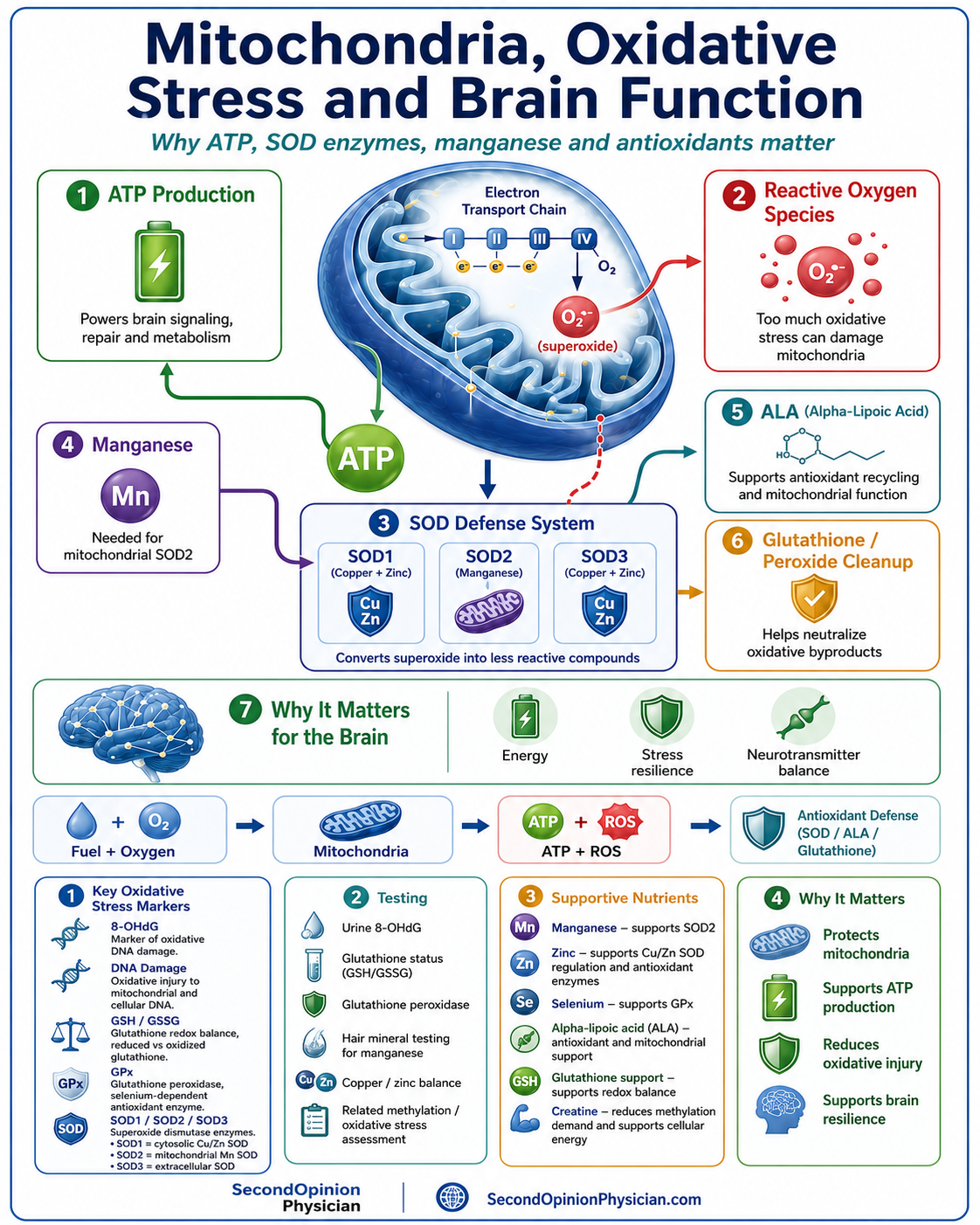
Mitochondria are specialized structures located in nearly every human cell. Their best-known function is producing adenosine triphosphate, or ATP, through oxidative phosphorylation. ATP is the immediate energy source used for electrical signaling, muscle contraction, active transport, protein synthesis, detoxification, methylation and cellular repair.
Mitochondria also help regulate calcium, heat production, steroid hormone synthesis, immune signaling, apoptosis, redox balance and the production or metabolism of several important cellular intermediates.
Energy production
Converts food-derived fuel and oxygen into ATP through the electron transport chain.
Redox regulation
Generates reactive oxygen species while also supporting antioxidant and repair systems.
Cell signaling
Influences calcium, inflammation, neurotransmission and cellular stress responses.
Cell survival
Helps determine whether a damaged cell repairs itself, adapts or undergoes programmed death.
How Mitochondria Produce ATP
Mitochondrial Disease Is Not the Same as Acquired Mitochondrial Dysfunction
Primary mitochondrial disease
Primary mitochondrial diseases are generally caused by pathogenic variants in mitochondrial DNA or nuclear genes that control mitochondrial structure and function. They may affect the brain, nerves, muscles, heart, liver, kidneys, hearing, vision, growth or endocrine system.
Secondary mitochondrial dysfunction
Acquired dysfunction may develop from oxidative stress, inflammation, nutrient deficiency, medication toxicity, toxic exposure, aging, metabolic disease, infection, poor sleep, inactivity or chronic illness. This does not automatically mean that the person has a genetic mitochondrial disorder.
Severe multisystem symptoms require specialist evaluation
Seizures, metabolic strokes, severe weakness, cardiomyopathy, developmental regression, exercise-induced rhabdomyolysis, lactic acidosis, major hearing or vision loss and strong maternal inheritance patterns may require neurological, metabolic or genetic evaluation.
Why Is the Brain Especially Vulnerable?
The brain has a very high energy requirement, stores little fuel and depends continuously on oxygen and glucose delivery. Neurons use ATP to maintain membrane potentials, release and recycle neurotransmitters, transport proteins along axons, maintain synapses and recover after each electrical signal.
Brain tissue is also rich in polyunsaturated lipids that can be damaged by oxidation. When mitochondrial energy falls and reactive oxygen species rise, the effects may include cognitive slowing, poor stress tolerance, sensory overload, sleep disturbance, irritability, impaired impulse control, psychosis vulnerability, fatigue and reduced rehabilitation capacity.
Superoxide, Nitric Oxide and Peroxynitrite
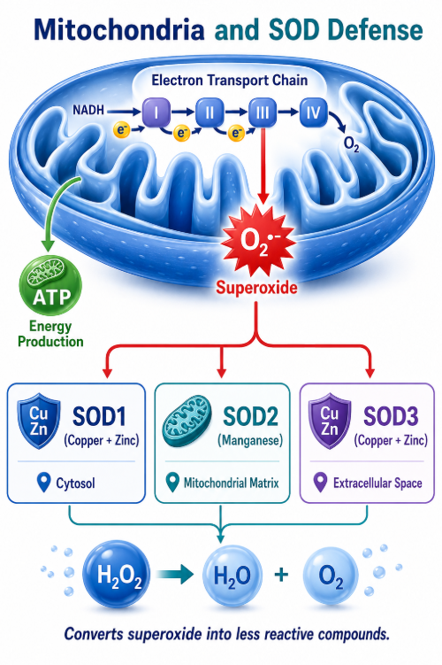
During normal respiration, a small percentage of electrons can escape the electron transport chain and reduce oxygen to superoxide. Superoxide is a reactive oxygen species that must be converted quickly into less reactive products.
From Superoxide to Oxidative and Nitrosative Injury
SOD1, SOD2 and SOD3: Three Superoxide Dismutase Systems
The SOD genes encode enzymes that convert superoxide into hydrogen peroxide. Hydrogen peroxide must then be cleared by catalase, glutathione peroxidase and related antioxidant systems. SOD activity without adequate downstream peroxide removal is incomplete protection.
| Enzyme | Main location | Mineral cofactors | Clinical relevance |
|---|---|---|---|
| SOD1 | Primarily cytosol; also mitochondrial intermembrane space | Copper and zinc | Controls intracellular superoxide outside the mitochondrial matrix and is sensitive to copper/zinc biology. |
| SOD2 | Mitochondrial matrix | Manganese | The principal superoxide defense inside mitochondria. |
| SOD3 | Extracellular matrix and vascular spaces | Copper and zinc | Helps regulate extracellular and vascular superoxide and preserve nitric-oxide signaling. |
Copper, Zinc and Manganese: Why Balance Matters
Copper
Copper is required for cytochrome-c oxidase in respiratory complex IV, SOD1, SOD3 and other enzymes. Poorly bound copper, however, can participate in oxidative reactions and damage lipids, proteins, DNA and mitochondria.
Zinc
Zinc helps stabilize SOD1 and SOD3, supports metallothionein and helps regulate copper. Low zinc may weaken antioxidant defense while increasing vulnerability to free-copper effects.
Manganese
Manganese is the metal cofactor for mitochondrial SOD2. Too little may reduce mitochondrial superoxide defense, but excess manganese is neurotoxic. It should be measured rather than supplemented indiscriminately.
Mitochondrial Dysfunction in Autism, Schizophrenia and Bipolar Disorder
Mitochondrial abnormalities and oxidative stress have been studied across several psychiatric and neurodevelopmental conditions. These findings do not mean that every patient has a primary mitochondrial disease. They suggest that cellular energy, antioxidant reserve and toxic burden may influence severity, treatment response and recovery.
Autism
Some autistic patients show oxidative stress, altered glutathione, mitochondrial abnormalities, developmental regression or poor tolerance of metabolic stress. Copper-zinc imbalance and SOD-related antioxidant capacity may add to the burden.
Schizophrenia
Research has identified oxidative and mitochondrial abnormalities in some patients. Smoking, antipsychotic metabolic effects, inflammation, poor diet, substance use and copper-zinc imbalance may increase mitochondrial stress.
Borderline personality disorder
BPD is not a mitochondrial diagnosis, and psychotherapy remains the core treatment. Severe emotional reactivity, sleep loss, trauma physiology, impulsivity, inflammation, substance exposure, nutrient deficiency and oxidative burden may interact with brain energy and stress tolerance.
The practical implication is to evaluate medication, sleep, nutrition, metabolic health, substance exposure, inflammation, copper-zinc balance, manganese, antioxidant reserve, methylation and overall function rather than reducing the case to one diagnosis.
How Mitochondrial Function Supports the Methylation Pathway
Methylation is often discussed as though it were only a folate and vitamin B12 pathway. In reality, methylation depends on cellular energy, mitochondrial one-carbon metabolism, amino-acid handling, redox balance and the removal of homocysteine and adenosine.
Methionine to SAM requires ATP
Methionine adenosyltransferase combines methionine with ATP to produce SAM. Poor ATP production may limit SAM generation even when methionine is available.
Mitochondria supply one-carbon units
Mitochondrial folate metabolism helps generate formate and other one-carbon units that support cytosolic nucleotide synthesis and methyl-group metabolism.
Homocysteine has two major exits
Homocysteine can be remethylated to methionine or directed through transsulfuration toward cysteine and glutathione. Energy, B12, folate, betaine, B6, zinc, protein and redox status influence these routes.
Adenosine must be metabolized
SAH hydrolase catalyzes a reversible reaction between SAH and homocysteine plus adenosine. If adenosine or homocysteine accumulates, the reaction may favor re-formation of SAH, which inhibits methyltransferases.
Mitochondrial Dysfunction Can Restrict Methylation at Several Points
Why Do We Use Creatine?
Creatine supports the phosphocreatine system, which rapidly buffers ATP in the brain and muscle. It may also reduce methylation demand because the body’s own creatine synthesis consumes a substantial amount of SAM-derived methyl groups.
ATP buffering
Phosphocreatine helps regenerate ATP during periods of high demand, supporting brain signaling, muscle function and recovery.
Methyl-group sparing
Supplemental creatine can reduce endogenous creatine synthesis and therefore lower one major use of SAM-derived methyl groups.
Not a complete solution
Creatine does not directly remove SAH, homocysteine or adenosine. It reduces one source of methylation demand while the underlying bottlenecks still require evaluation.
Creatine requires clinical context
Creatine can increase serum creatinine without necessarily causing kidney damage. Kidney function, hydration, medications, cystatin C and the overall clinical picture may be important when interpreting follow-up results.
Which Tests Help Evaluate Mitochondrial and Oxidative Stress?
No single test proves acquired mitochondrial dysfunction. The most useful strategy combines symptoms, history, medications, nutrient status, oxidative-damage markers, antioxidant reserve and the clinical response over time.
| Marker | What it may show | Limitation |
|---|---|---|
| 8-OHdG | Oxidative damage to DNA and the balance between injury and repair | Can rise from many causes and does not identify the source alone |
| Glutathione | Reduced antioxidant reserve or altered redox balance | Interpretation depends on whether reduced, oxidized or total glutathione is measured |
| Glutathione peroxidase | Peroxide-removal capacity and selenium-dependent antioxidant function | Influenced by selenium, inflammation, genetics and sample type |
| SOD activity | Superoxide-dismutase antioxidant capacity | May not distinguish SOD1, SOD2 and SOD3 or identify the deficient cofactor |
| Copper, ceruloplasmin and zinc | Copper binding, estimated free copper and zinc balance | Inflammation, albumin, hormones and supplements affect interpretation |
| Whole-blood manganese | Current manganese status or exposure pattern | Both deficiency and excess matter |
| Hair elements | Longer-term exposure pattern for manganese and toxic metals | Hair treatment and contamination can distort results |
| SAM, SAH, methionine and homocysteine | Methyl-donor supply, inhibition and pathway congestion | Abnormal values do not prove mitochondrial dysfunction alone |
Treating Mitochondrial Dysfunction: A Layered Strategy
Treatment depends on whether the problem is a primary mitochondrial disease, acquired dysfunction or one component of a broader psychiatric, neurological or metabolic illness. The goal is not to give every patient the same “mitochondrial cocktail.”
1. Remove ongoing stressors
- Address smoking, alcohol, cannabis or stimulant exposure
- Review medications that may affect mitochondria
- Treat sleep apnea and chronic sleep loss
- Investigate credible toxic exposures
2. Correct measured deficiencies
- Protein and amino-acid adequacy
- Zinc, manganese, magnesium and selenium when deficient
- Vitamin D, B12 and other documented deficiencies
- Copper correction according to copper, zinc and ceruloplasmin
3. Support energy and redox balance
- Creatine for ATP buffering and methyl-group sparing
- CoQ10 when clinically appropriate
- NAC, glycine or glutathione support according to tolerance
- Alpha-lipoic acid and vitamins C/E selectively
4. Build mitochondrial capacity
- Gradual aerobic and resistance exercise
- Physical rehabilitation when deconditioned
- Stable glucose and adequate calorie intake
- Ketogenic strategies only in selected patients
5. Address methylation bottlenecks
- Distinguish low SAM from high SAH
- Review homocysteine, adenosine-related metabolism and kidney function
- Support transsulfuration and glutathione when appropriate
- Avoid automatic folate or methyl-donor treatment
6. Monitor the whole patient
- Psychiatric stability and medication adherence
- Sleep, cognition, energy and exercise tolerance
- Weight, glucose, lipids and blood pressure
- Repeat biomarkers only when they will change treatment